Tower Imaging’s Dr. Anjali Date Discusses Henry Mayo’s Sheila R. Veloz Breast Center for October’s Breast Cancer Awareness Month

Tower Imaging’s Dr. Anjali Date Discusses Henry Mayo’s Sheila R. Veloz Breast Center for October’s Breast Cancer Awareness Month

Henry Mayo Newhall Hospital’s Sheila R. Veloz Breast Center is recognized as a Breast Imaging Center of Excellence by the American College of Radiology, and was created through generous philanthropy and the experiences of breast cancer survivors, patients and their families. “What makes the Sheila R. Veloz Breast Imaging Center so important is that it is a community center: we are founded by the community, funded by the community, staffed with members of our community and we serve the women in our community,” said Anjali Date, MD, Lead Interpreting Physician.

There are many options for breast care, but the Sheila R. Veloz Breast Center is unique in that it provides excellent care with a genuine kindness and compassion to ensure patients the best possible outcome. “Not only do we perform the most up-to-date, state of the art Breast Imaging available, but we also provide high-risk genetic screening and counseling in conjunction with Ambry Genetics,” said Dr. Date. “This means we can help identify women who would benefit from additional methods of screening for breast cancer such as annual MRI, tailoring our approach to how best to serve each woman of Santa Clarita as individuals.”

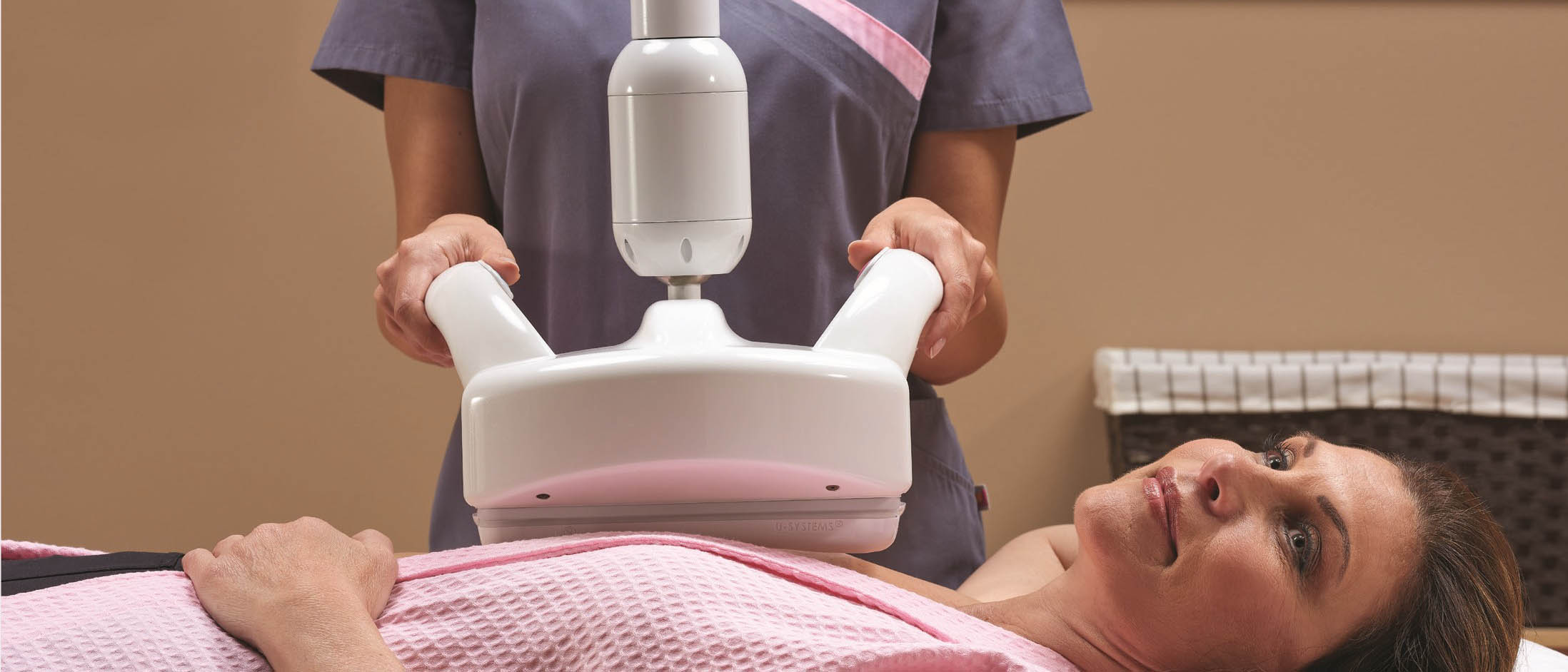
Our Sheila R. Veloz Breast Center features the latest in digital mammography and ultrasound detection. The new software in our Hologic 3D quorum digital mammography machines provides higher resolution imaging that identifies cancer sooner and clearer. It also improves reading time which results in faster more accurate results. “With the aid of advanced technology, fewer false positives and improved cancer detection means that we can find cancer earlier which greatly improves overall survival,” said Dr. Date.


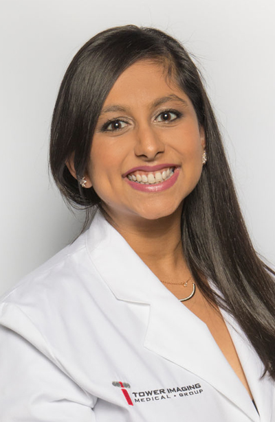
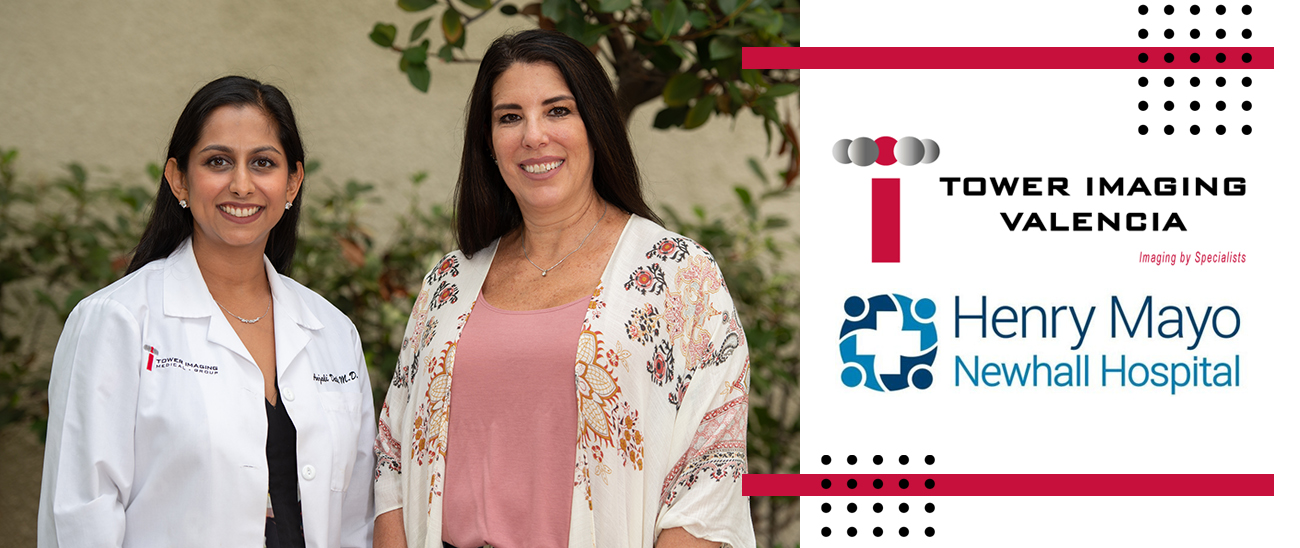
Anjali Date, MD, Lead Interpreting Physician, Shelia R. Veloz Breast Center

Later this year, our breast center will be acquiring the SCOUT Radar Localization system. The SCOUT system enables surgeons to precisely target the affected tissue to pinpoint its location within 1mm, which can mean more successful surgeries, optimized breast conservation strategies, and enhanced outcomes for women.

October is National Breast Cancer Awareness Month and Dr. Date shared that it’s one of her favorite months in the Santa Clarita Valley. “Being part of the Sheila R. Veloz Breast Center, I have been given opportunities to speak and educate the women of our community on breast health, screening, and cancer prevention,” said Dr. Date. “With COVID shutting down our facilities for two months March and April of 2020, and the lingering hesitancy to get back to our routine health care appointments, we have seen a significant decrease in the number of screening mammograms, and subsequently and increase in the number of larger, later stage breast cancers. My drive and desire to help these women and the rest of our community resonates now more than ever: early detection is the best prevention.”

“I have immense pride in the work I do here, and I am reminded on a daily basis of how important it is for women to receive quality healthcare and annual screening for breast cancer,” said Dr. Date. In honor of Breast Cancer Awareness Month, the team at Henry Mayo’s Sheila R. Veloz Breast Center would like to encourage the community to schedule annual mammogram screenings now because early detection is key to saving lives! For more information and to schedule an appointment, please call 661-200-1099 or visit, https://www.henrymayo.com/our-services/breast-center/

Thank you for the continued support that enables the Sheila R. Veloz Breast Center to provide state-of-the-art healthcare for the Santa Clarita Valley and surrounding communities. In honor of Breast Cancer Awareness Month, we ask that you please consider a gift.

TSJI Conducts Successful Holiday Toy Drive for Kids

Tower Saint John’s Imaging would like to thank all those who donated to our Holiday Toy Drive. We are all so humbled by your donations. Your support has repeatedly played a key role in our success. There is no way to fully express our gratitude for all that participated. We are continually inspired by the dedication and generosity of donors like yourselves who answer the call to give again and again. Your amazing gifts went to the following charities:

Jewish Family Services – Children from Domestic Violence Families
Impacto – After school program for kids
Ride for Kids – Pediatric Brain Tumor Foundation
Chai Life – Pediatric Illness
Maryvalle – Teenage Foster Girls
Maharlika Lions Club – Children in Hospital In-Patient Care

Thank you again for your support and HAPPY HOLIDAYS!

Introducing the SmartCurve™ System

The More Accurate1-6 Genius™ Exam Just Got More Comfortable7

Mammograms are a woman’s best defense in detecting breast cancer early, but we know that the fear of pain causes many women to delay scheduling their annual mammogram.

That’s why we proudly offer the Genius™ 3D Mammography™ exam that combines better, earlier breast cancer detection1-6 with the improved comfort of the SmartCurve™ breast stabilization system.

Welcome to a mammogram that is more accurate1-6 and more comfortable.7

What to Expect During your Exam

Getting a mammogram with the SmartCurve™ system is just like getting a regular Genius™ exam – only more comfortable!7

The curved design of the compression device mirrors the shape of a woman’s breast to reduce pinching and applies uniform compression over the entire breast for added comfort.

The technologist will view the images of your breasts at the computer workstation to ensure quality images have been captured for review. A radiologist will then examine the images and report results to either your physician or directly to you.

Ask about getting your Genius™ exam with the SmartCurve™ system by calling us at 310.264.9000!

The Genius™ 3D Mammography™ exam (a.k.a. Genius™ exam) is acquired on the Hologic 3D Mammography™ system and consists of a 2D and 3D™ image set, where the 2D image can be either an acquired 2D image or a 2D image generated from the 3D™ image set. The Genius™ exam is only available on the Hologic 3D Mammography™ system. Please consult your physician for a full list of benefits and risks associated with mammography. PP-01979 Rev. 001 ©2018 Hologic, Inc. Hologic, 3D, 3D Mammography, Dimensions, Genius, Selenia, and associated logos are trademarks and/or registered trademarks of Hologic, Inc. and/or its subsidiaries in the United States and/or other countries. 1. FDA submissions P080003, P080003/ S001,P080003/S004, P080003/S005. 2. Friedewald SM, Rafferty EA, Rose SL, et al. Breast cancer screening using tomosynthesis in combination with digital mammography. JAMA. 2014 Jun 25;311(24):2499-507. 3. Zuckerman SP, Conant EF, Keller BM, et al. Implementation of Synthesized Two-dimensional Mammography in a population-based Digital Breast Tomosynthesis Screening Program. Radiology. 2016 Dec;281(3):730–736. 4. Skaane P, Bandos A, Eben EB, et al. Two-view digital breast tomosynthesis screening with synthetically reconstructed projection images: comparison with digital breast tomosynthesis with full-field digital mammographic images.

Radiology. 2014 Jun;271(3):655–63. 5. Bernardi D, Macaskill P, Pellegrini M, et. al. Breast cancer screening with tomosynthesis (3D mammography) with acquired or synthetic 2D mammography compared with 2D mammography alone (STORM-2): a population-based prospective study. Lancet Oncol. 2016 Aug;17(8):1105–13. 6. McDonald ES, Oustimov A, Weinstein SP, et al. Effectiveness of Digital Breast Tomosynthesis Compared With Digital Mammography: Outcomes Analysis From 3 Years of Breast Cancer Screening. JAMA Oncol. 2016 Jun 1;2(6):737–43. 7. Smith, A. Improving Patient Comfort in Mammography. Hologic WP-00119 Rev 003 (2017).

October is Breast Cancer Awareness Month

October is Breast Cancer Awareness Month, so do something important for those you love by doing something for your health. Schedule your exam today.

Breast Cancer is the second most common type of cancer for women in the United States. Whether or not you have been yet, you will be exposed to breast cancer at some point in your life. At least 1 in every 8 women will be diagnosed with it during their life. Breast screenings can save your life by detecting cancer at its earliest and most treatable stage.

Here at Tower Saint John’s Imaging, we offer the most advanced
Breast Imaging Services which include:

  • 3D Mammography (Digital Breast Tomosynthesis)
  • Automated Breast Ultrasound (ABUS)
  • Breast MRI
  • MRI-guided biopsy

Schedule your exam today by calling:
(310)264-9000

Invenia ABUS 2.0: The Only FDA-Approved Ultrasound Technology for Breast Cancer Detection in Women with Dense Breast Tissue

The Dense Breast Dilemma

Over 40 percent of women have dense breast tissue, one of the strongest common risk factors for developing breast cancer. In dense breasts, cancers may be masked on 2D mammography – potentially delaying diagnosis in these women. Both dense breast tissue and cancer appear white on a mammogram, creating a dangerous camouflage effect and a dilemma for radiologists whose goal is to find breast cancer as early as possible.

Detecting More Cancer

GE Healthcare offers the only FDA-approved ultrasound supplemental screening technology that is specifically designed for detecting cancer in dense breast tissue. Compared to mammography alone, Invenia ABUS 2.0 imaging looks differently at dense breast tissue, providing a comprehensive view of the breast. This technology has demonstrated a 35.7% increase in cancer detection over mammography alone for these women.

Supplemental imaging with Invenia ABUS 2.0 transforms breast care from reactive to proactive. Clinical research studies demonstrate that when used as an adjunct to mammography, small cancers visible only through ABUS were predominantly invasive and node-negative. Detecting them at this earlier stage has important prognostic implications and can reduce the cost of care.

Learn More about Abus 2.0

Contact Tower Saint John’s Imaging to learn more about Automated Breast Ultrasound technology and how it can help save lives.

Tower Saint John’s Imaging Toy Drive Was a Success!

Tower Saint John’s Imaging conducted their successful Toy Drive benefiting the following organizations:

  • Salvation Army with it’s collaboration with Toys for Tots, an official mission of the Marine Corps Reserve.
  • Jewish Family Services of Los Angeles, a multi-service Agency who’s goals are to strengthen and preserve individual, family and community life.
  • Chai Lifeline, they bring joy to the lives of seriously ill children and their families.

The Power of Combining ABUS and Tomosynthesis to Find Small Cancers

40% of women have dense breast tissue and for these women, mammography screenings alone may not be enough to find breast cancer. Both cancer and dense tissue appear white on a mammogram, enabling cancer to hide.

Speaking on the benefits of utilizing Automated Breast Ultrasound to help detect cancer in dense breasts, Tower Imaging Medical Group’s Dr. Daniel Kirsch was quoted as saying, “We implement ABUS because it offers a true advance in screening ultrasound evaluation of the breast.”

Dr. Kirsch further elaborated by stating, “Studies have shown that tomosynthesis can improve cancer detection, and decrease recall rates or false positives. With the ability to see through overlapping tissue, tomosynthesis also provides improved localization and characterization of masses,” said Arnold Honick, MD, Radiologist, RCI, PLC, Radiology Consultants of Iowa in Cedar Rapids, Iowa. “However, tomosynthesis is still X-ray based and sensitivity is reduced in women with extremely dense breasts, which means that some masses will still be masked and likely not detected. This is where ABUS, utilizing a different imaging technology, is complementary to mammography and can improve overall screen-detected breast cancer.”

To view more information about Automated Breast Ultrasound, and read more about Dr. Kirsch’s statement of benefits, view the full article on Aunt Minnie’s site here: https://www.auntminnie.com/index.aspx?sec=eba&sub=eml&pag=dis&itemId=121950&muid=10866708

Contact Tower Saint John’s Imaging at 310.264.9000 to schedule an ABUS exam.

Go Big or Go Bust? How Less-than-Large Radiology Practices Can Survive the Consolidation Era

Speaking on the ever-challenging environment in the radiology services industry, Tower Imaging Group’s Dr. Bruce Yawitz was featured and quoted in www.radiologybusiness.com discussing how radiology facilities can adapt and survive the present day’s many business challenges.

Speaking on Tower Imaging’s strategical objectives, he was quoted as saying, “We pride ourselves on being a specialty-based, small boutique group, but these days I don’t think that’s enough,” says Bruce Yawitz, MD, president of Tower Imaging. “We need to have a bigger footprint, and sharing assets with other like-minded groups under the umbrella of Strategic Radiology will provide everything that’s important to our hospital partners, while allowing us to maintain the independence we’ve always cherished.”

To view the complete article, please go to the following link: Go Big or Go Bust? How Less-than-Large Radiology Practices Can Survive the Consolidation Era

Improved Diagnostic Confidence Now Available at Tower Saint John’s Imaging with the Discovery MI DR PET/CT

This innovative PET/CT imaging system from GE Healthcare may help you increase your diagnostic confidence and enhance the care of your patients. The Discovery MI DR PET/CT uses advanced detector technology and compared to previous scanners, can deliver:

  • High PET sensitivity and count rate performance to image all tracers *2 mm resolution over the entire FOV with SharpIR
  • Up to 82 percent reduction in CT dose with ASiR, at the same image quality
  • Up to 2X improvement in quantitative accuracy (SUVmean) and image quality (SNR) with Q.Clear
  • Platform compatible with advanced digital solutions designed to connect machines, people and data through a portfolio of healthcare analytics applications.

The Discovery™ MI DR PET/CT imaging system from GE Healthcare uses revolutionary LightBurst detector technology to improve scanning speed and diagnostic accuracy.

In addition to faster scanning times, this scanner will allow lower radiation dosing with new state-of-the-art LYSO detectors. Discovery MI DR was created to help clinicians diagnose and stage disease earlier and better guide treatment strategies while enabling more compelling research with more novel, faster decaying tracers. This system also features the latest diagnostic CT innovations to provide a “one-stop-shop” diagnostic experience.

With the Discovery MI DR system, we continue to raise the level of excellence in healthcare in our area. We are proud to be on the leading edge of advancing the quality of cancer and cardiac care for our local, regional and international patients.

See how this remarkably advanced PET/CT system can help make a difference in patient care by calling us at 310.264.9000 and requesting more info!

Publication: Preoperative Ultrasound Assessment of Regional Lymph Nodes in Melanoma Patients Does Not Provide Reliable Nodal Staging: Results From a Large Multicenter Trial

John F. Thompson, MD,*1 Lauren E. Haydu, PhD,1 Roger F. Uren, MD1, Robert H. Andtbacka, MD2, Jonathan S. Zager, MD3, Peter D. Beitsch, MD4, Doreen Agnese, MD5, Nicola Mozzillo, MD6, Alessandro Testori, MD7, Tawnya L. Bowles, MD8, Harald J. Hoekstra, MD, PhD9, Mark C. Kelley, MD10, Jeffrey Sussman, MD11, Schlomo Schneebaum, MD12, B. Mark Smithers, MBBS13, Gregory McKinnon, MD14, Eddy Hsueh, MD15, Lisa Jacobs, MD16, Erwin Schultz, MD17, Douglas Reintgen, MD3, John M. Kane, MD18, Erica B. Friedman, MD1, Hejing Wang, MD19, Lisa Van Kreuningen, MS20, Vicki Schiller, MD21, David A. Elashoff, PhD19, Robert Elashoff, MD19, Alistair J. Cochran, MD19, Stacey Stern, MS20, Mark B Faries MD20, and the MSLT-II Trial Group

1Melanoma Institute Australia, The University of Sydney, 2Huntsman Cancer Institute, 3H. Lee Moffitt Cancer Center, 4Dallas Surgical Group, 5Ohio State University, 6Istituto Nazionale dei Tumori Napoli, 7Istituto Europeo de Oncologia, 8IHC Cancer Services – Intermountain Medical Center, 9University Medical Center Groningen, 10Vanderbilt University, 11University of Cincinnati, 12Tel-Aviv Sourasky Medical Center, 13Princess Alexandra Hospital, 14Tom Baker Cancer Centre, 15Saint Louis University, 16Johns Hopkins Medical Institute, 17City Hospital of Nürnberg, Paracelsus Medical University, 18Roswell Park Comprehensive Cancer Center, 19University of California at Los Angeles; 20John Wayne Cancer Institute, 21Providence St John’s Health Center

Corresponding author:
John F Thompson MD
Melanoma Institute Australia
40 Rocklands Road
NORTH SYDNEY NSW 2060
AUSTRALIA
Tel: +612 9911 7366

E:  john.thompson@melanoma.org.au

ABSTRACT

Introduction: It has been suggested that preoperative ultrasound (US) assessment of regional lymph nodes in patients who present with primary cutaneous melanoma provides accurate staging and avoids the need for sentinel node (SN) biopsy.  However, in most single institution studies the reported sensitivity for preoperative US detection of nodal metastases has been low.

Methods: Preoperative US data and SNB results were analyzed for patients enrolled at 20 centers participating in the screening phase of the second Multicenter Selective Lymphadenectomy Trial.  Excised SNs were histopathologically assessed and considered positive if any melanoma was seen.

Results: SNs were identified and removed from 2786 patients who had pre-operative US evaluation.  Among those patients, 531 had SN metastases. US was positive (abnormal) in 85 patients (3.1%). Among SN-positive patients, 39 (7.3%) had an abnormal US. When analyzed by lymph node basin, 3222 basins were evaluated, and 38 were true positive (1.2%). By basin, the sensitivity of US was 6.8% (95% CI: 4.7%-8.9%) and the specificity 98.0% (95% CI: 97.5%-98.5%).  Median cross sectional area of all SN metastases was 0.13mm2; in US true-positive nodes it was 4.8mm2. US sensitivity increased with increasing Breslow thickness of the primary melanoma (0% for ≤1mm thickness, 12.4% for >4mm thickness). US sensitivity was not significantly greater with higher trial center volume or with pre-US lymphoscintigraphy.

Conclusion: In the MSLT-II screening phase population, SN tumor volume was usually too small to be reliably detected by US. For accurate nodal staging to guide the management of melanoma patients, US is not an effective substitute for SN biopsy.


Next Page »
Tower Saint John's Imaging is an ACR accredited facility. Call 310.264.9000 or Click to schedule appointment.SCHEDULE APPOINTMENT